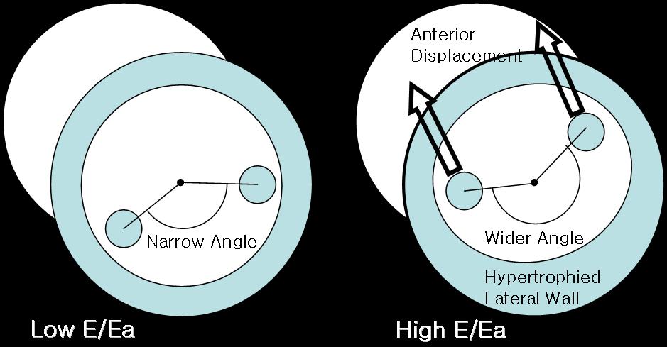
BACKGROUND Various anomalies of papillary muscles (PM) have been shown to be associated with apical hypertrophic cardiomyopathy (ApHCM). We sought to investigate whether various changes of PM are associated with diastolic function in ApHCM. METHODS Twenty-three ApHCM patients (14 male; mean age: 66) underwent echo-Doppler evaluations. In end-diastolic phase, the 16 LV segmental thicknesses and PMs thicknesses were measured in apical-view. The distance and angle between both PM were assessed in short-axis view at the mid LV level. RESULTS Early diastolic mitral inflow velocity/septal mitral annular velocity (E/Ea) was significantly correlated with the left atrial volume index (36.9ТБ16.4mL, r=0.331, p=0.037), the number of hypertrophied (> 12 mm) segments (3.8ТБ2.8, r=0.485, p=0.002). Interestingly the angle between both PM is significantly correlated with E/Ea (183.9ТБ27.6ТА, r=0.378, p=0.023). Wider angle between both PM means anterior displacement of PM (Figure). But, the thickness of maximal hypertrophied segment of LV (18.5ТБ0.4mm, r=0.231, p=0.125), the maximal thickness of PM (9.8ТБ1.8mm, r=0.127, p=0.413) and the distance between both PMs (24.6ТБ4.9mm, r=0.246, p=0.110) were not statically correlated with E/Ea. CONCLUSION the angle between both PM, suggesting PM displacement, is associated with diastolic function of LV in ApHCM.
|