Background: Clinical implications of the initial echocardiographic characteristics of pericardial effusion (PE) have not been clearly demonstrated.
Methods: Echocardiographic evaluation was performed in consecutive 178 patients with moderate to severe PE, and the etiology of PE was determined by clinical and pathologic studies. Echocardiography was repeated 46ТБ56 weeks.
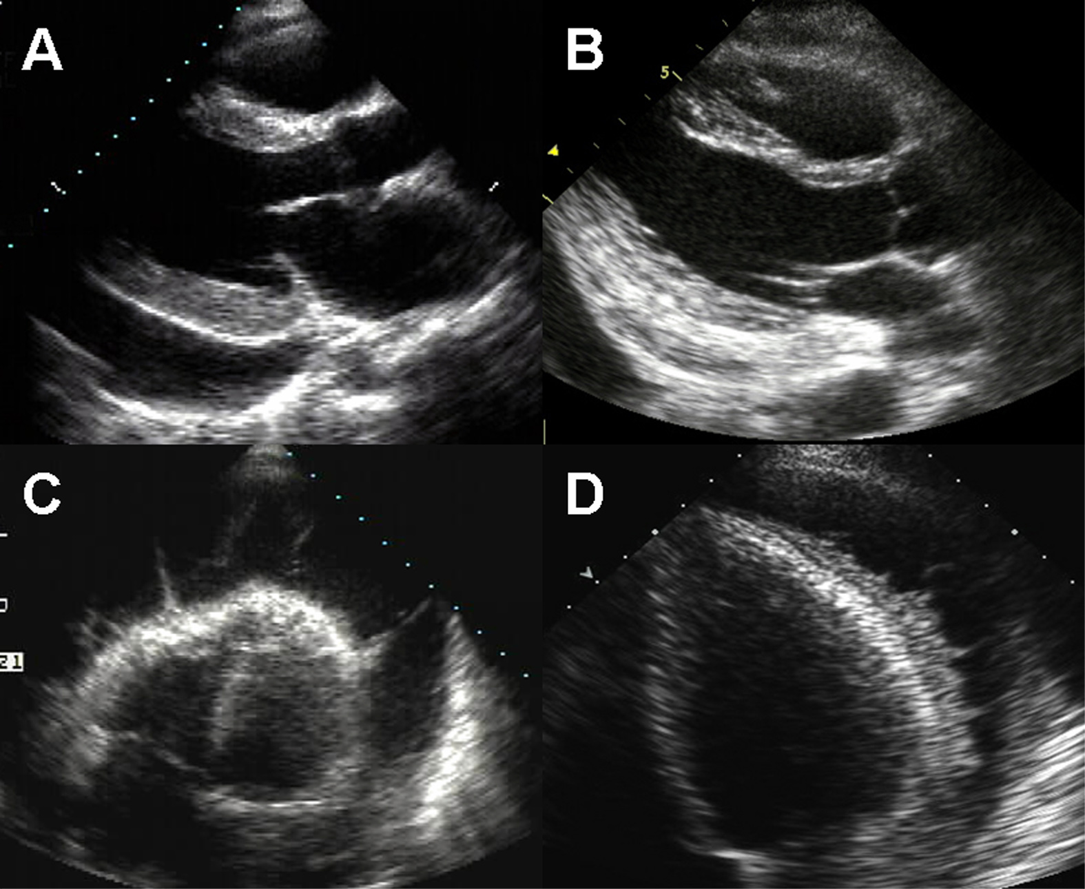
Results: Echo-free PE was shown in 142 patients (80%, group A, Fig A), while echogenic PE was found in 36 patients (20%, group B). In group B, diffuse echogenic PE (Fig B) was shown in 7 patients and intrapericardial fibrinous strands (Fig C) and/or echogenic frond-like materials (Fig D) were detected in 29 patients. The prevalence of echogenic PE was the highest in tuberculosis (56.3%). All 49 patients with uremia or congestive heart failure showed clear echo-free PE. During the follow-up, the incidence of constrictive pericarditis and recurrent PE were highest in malignancy (38.6%) and tuberculosis (31.3%), respectively. The incidence of constrictive pericarditis (3.5 vs. 27.8%, p<0.001) and recurrent PE (9.2 vs. 22.2%, p<0.05) were significantly lower in group A than group B. The echogenic PE was the only independent predictor of the events by multiple stepwise logistic regression analysis (p<0.05), regardless of the PE etiology.
Conclusion: Echogenic materials in PE predict pericardial complications such as recurrence and constrictive pericarditis, irrespective of underlying diseases.
|