Introduction: Pulmonary vein (PV) antrum has been accepted as the principal targets in atrial fibrillation (AF) ablation. It is difficult to know the true anatomical location of the PV antrum with fluoroscopy only. Even it will not be easy with electroanatomical mapping system unless image integration is available. Intracardiac echocardiography (ICE) is useful in anatomical landmark detection as well as facilitating transseptal puncture and RF ablation monitoring. This study was designed to evaluate the gap between electrogram-based PV os determination versus ICE-guided PV antrum determination.
Methods: Ten patients with AF who underwent PV antrum isolation were evaluated. PV antrum was identified using ICE. Electrogram-based PV os was determined by the most proximal point with atrial electrogram amplitude < 0.5 mV. The distance between the PV antrum border and PV os was measured on ICE system or electroanatomical mapping system. Three sides of each PV were evaluated: superior, anterior, and posterior sides for the superior PVs and inferior, anterior and posterior sides for the inferior PVs.
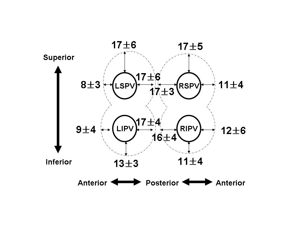
Results: Measured distances are plotted on the figure (unit: mm).
Conclusion: Discrepancy was especially large in superior and posterior sides. Therefore these regions would need more wide-area ablation for PV antrum isolation.
|