| ЙпЧЅЧќНФ : ЦїНКХЭ
|
СЂМіЙјШЃ - 510644 45 |
| Intravascular Ultrasound Observation of Neointimal Distribution for Single Long (23mm~33mm) Drug-Eluting Stents: Comparison of SES and PES |
| ъГыЊ
ыэъЕ ыьАьыЃь ьЌьЅыДъГМ |
| ьЁАьЄъВН, эьЙэИ, ь ыэ, ъЙыГДы, ьЄь ьЇ, ь ьыЏИ, ьДьЇьД, ыЈьАНьБ, ъЙэь, эьБьБ, ъЙьЄы
, ъЙъЖыАА |
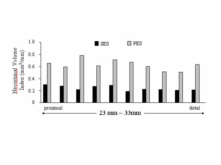
Background:Previous IVUS studies have reported that different types of drug-eluting stents showed different patterns of neointimal hyperplasia (NIH) distribution within stent. However, these patterns were limited to relatively short stent (<20 mm). The aim of this study is to evaluate NIH distribution within single long (23 mm to 33 mm) stent.
Methods:From our database, 36 patients with sirolimus-eluting stents (SES: 23 mm, 28 mm or 33mm) and 13 patients with paclitaxel-eluting stents (PES: 24mm, 28mm or 32 mm), who underwent serial (baseline, follow-up: 8.2ТБ2.0 months) IVUS examination, were enrolled in this study. All patients were treated with single stent. Every stent was divided into 10 subsegments for evaluation of NIH amount.
Results:Overall, neointimal volume index (volume/length) was significantly lower in SES than PES (3.49ТБ5.06 mm3/mm vs. 8.52ТБ7.08 mm3/mm; p=0.009). The SES showed a significantly lower or a trend toward a lower amount of NIH than PES in each subsegment. There was significantly higher amount of NIH at proximal edge compared to mid-body (p<0.05), whereas the PES did not show different amount of NIH in each subsegment (p=NS).
Conclusion:In single long stent, the PES showed greater NIH amount throughout entire stent length than SES. There was a different pattern of NIH distribution between PES and SES, which was comparable with previous reported distribution pattern in each stent.
|
|
|
Warning: getimagesize(/home/virtual/circulationadmin/htdocs/econgress/conference/abstract/img_files/DEScompleslesion.jpg) [function.getimagesize]: failed to open stream: No such file or directory in /home/virtual/circulationadmin/new/econgress/conference/manage/schedule/view_abstract.php on line 164
|
|