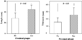
Purpose: In this study, we used multidetector computed tomography (MDCT) to detect myocardial bridge and evaluate the anatomical properties of it. Methods: 64-slice MDCT was conducted on 666 patients suspected with coronary artery disease. Myocardial bridge was diagnosed when an intramural segment of a coronary artery was visualized on axial and multiplanar reconstruction (MPR) images. Results: Among the 666 patients, 38 patients(6 %) were found to have myocardial bridge. In nineteen patients (50%), myocardial bridge was located in mid left anterior descending artery (LAD). And in thirteen patients (34%), the MB was located in the proximal or distal LAD. The length of tunneled artery was mean 16у, from 6.9у to 30у. And the maximum thickness of myocardial tissue was between 0.5уand 3.9у, with mean of 1.8у. The length of MB was significantly correlated with their thickness (p < 0.05). Although most of intramural segment was free of coronary wall lesions, arterial segment immediately proximal to the myocardial bridge had atherosclerotic plaque in 10 of 38 cases (26%). The presence of atherosclerotic plaque in the proximal to the myocardial bridge correlated with the thickness and length (p < 0.05). Conclusions: In our study, we described the incidence of the myocardial bridge and its anatomical properties with MDCT. The MDCT is a useful and noninvasive tool for evaluating myocardial bridge.
|