| єя«•«ьљƒ : ∆чљЇ≈Ќ
|
ЅҐЉцєш»£ - 540152 276 |
| Image Analysis-based Lung and Heart Region Detection and Diagnosis
Support System for Chest X-ray of Patient with Dyspnea |
| к≥Дл™ЕлМАнХЩкµР мГЭм≤ім†Хл≥ікЄ∞мИ†к∞Ьл∞Ь мВђмЧЕлЛ®¬є, к≥Дл™ЕлМАнХЩкµР мЭШк≥ЉлМАнХЩ мЭШмЪ©к≥µнХЩк≥Љ¬≤, к≥Дл™ЕлМАнХЩкµР мЭШк≥ЉлМАнХЩ мЭШл£Мм†Хл≥інХЩкµРмЛ§¬≥, к≥Дл™ЕлМАнХЩкµР лПЩмВ∞мЭШл£МмЫР мЛђмЮ•лВік≥ЉвБі , к≥Дл™ЕлМАнХЩкµР лПЩмВ∞мЭШл£МмЫР нШЄнЭ°кЄ∞лВік≥Љ 5 |
| мДЬмДЭнГЬ¬є, л∞ХнЭђм§А¬≤, кєАлѓЉмИШ¬є, мЖРм∞љмЛЭ¬≥, л∞ХнШХмД≠вБі, м†ХмєШмШБ5, кєАмЬ§лЕД вБі |
Abstract
Objective: Chest X-ray images are the common and widely used in clinical practice. Many researches have been proposed to segment and analyze chest X-ray images. However interpretation of chest X-ray images is still challenging and interesting research field because of the complication and variety of the images. Therefore, in this study, we propose a diagnosis support system for chest X-ray image based on image analysis to evaluate normality.
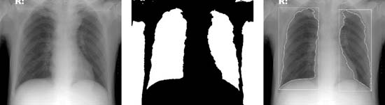
Methods: Various chest X-ray images with diagnosis by clinical experts were collected. To segment regions from the chest X-ray images, thresholding and morphological methods have been applied. Moreover measurement and texture analysis techniques were performed on the segmented region to evaluate normality.
Results: To show the effectiveness of the proposed method, we have applied 10 patients' images. Among the patients, 8 patients show abnormality in chest X-ray images, and 5 patients have cardiomegaly. From the experimental results, the proposed method based on image processing provided similar results with the clinical experts. The detection accuracies of cardiomeraly and overall normality of the proposed method were 0.9 and 0.8 respectively. Moreover the sensitivity and specificity for cardiomegaly detection were 0.8 and 1 respectively.
Conclusion: We proposed a diagnosis support system based on image processing to analyze chest X-ray images. Moreover the effectiveness of method was shown through experiments. However the method is limited to cardiomegaly and normality. Therefore the expended researches to analyze other characteristics such as nodules, blood vessels, and etc., and to apply other radiography images are required as further researches.
|
|
Table. Analysis Results
|
|
Accuracy |
Sensitivity |
Specificity |
|
Cardiomegaly |
0.9 |
0.8 |
1 |
|
Overall Normality |
0.8 |
0.5 |
0.875 |
|
Warning: getimagesize(/home/virtual/circulationadmin/renewal/econgress/conference/abstract/img_files/ExampleofImageAnalysisResult.jpg) [function.getimagesize]: failed to open stream: No such file or directory in /home/virtual/circulationadmin/new/econgress/conference/manage/schedule/view_abstract.php on line 164
|
|


