| л∞ХкЈЬнЩШ¬є, нЩНкЈЄл£®¬є , мЭіл≥ік≤љ¬є, мЭімГБнЭђ¬є , л∞ХмҐЕмД†¬є , мЛ†лПЩкµђ¬є , кєАмШБм°∞¬є , мЛђліЙмД≠¬є , мµЬм†ХнШД¬≤ |
Background: Prediction of left ventricular (LV) apical thrombus formation in patients with LV dysfunction is important for long-term mortality and morbidity. However, conventional echo-Doppler parameters including EF could not predict LV thrombus formation well. The LV vortex flow may play a important role in thrombus formation in the LV apex. The aim of this study was to assess clinical usefulness of the LV vortex flow analysis for predicting apical thrombus in patients with LV dysfunction.
Methods: Thirty patients with LV dysfunction (LVEF < 40%) underwent 2-D contrast echocardiography(CE) with intravenous infusion of Definity¬Ѓ (Lantheus Medical Imaging, Inc. North Billerica, MA) and imaged at an mechanical index of 0.4-0.6 in the A4C and APLX views. Study population consisted of two groups, thrombus group: patients with apical thrombus (n = 12) and non-thrombus group: without apical thrombus (n = 18). Location and pulsatility parameters of the LV vortex were measured using Omega flow¬Ѓ (Siemens Medical Solutions, Mountain View, CA) and compared between two groups.
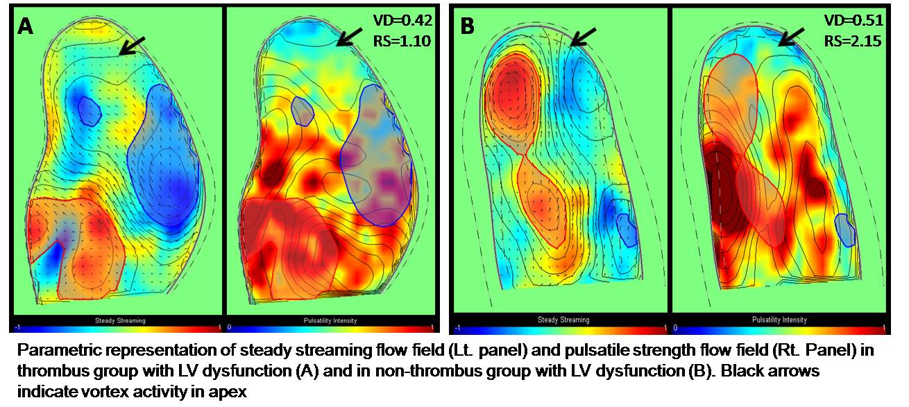
Results: LVEF, LV mass index, and other echocardiographic findings were not significantly different between two groups. In non-thrombus group, a coherent vortex was placed near the LV apex. However, in thrombus group, vortex was located in the center of the LV, and did not extend to the LV apex. In thrombus group, vortex parameters such as vortex depth (0.429 ± 0.108 vs. 0.531 ± 0.082, p = 0.018) and relative strength (1.418 ± 0.270 vs. 1.850 ± 0.399, p = 0.014) were significantly lower than non-thrombus group. Figure shows parametric representations of steady streaming flow field (left panel) and the pulsatile strength field (right panel) in thrombus (A) and non-thrombus (B) group.
Conclusions: LV vortex flow analysis is clinically useful for predicting the LV apical thrombus in patients with LV dysfunction, which offers a new method to application for anticoagulation treatment in patients with LV dysfunction.
|