| єя«•«ьљƒ : ±Єњђ
|
ЅҐЉцєш»£ - 540213 234 |
| Virtual Dissection CT Angiography with Endocardiovascular Volume Rendering: a New Three-Dimensional Imaging for Intracardiac Anomaly in Congenital Heart Disease |
| мДЄмҐЕл≥СмЫР мШБмГБмЭШнХЩк≥Љ¬є, мЖМмХДм≤≠мЖМлЕДк≥Љ ¬≤ , нЭЙлґАмЩЄк≥Љ¬≥ |
| кєАмЦСлѓЉ¬є, , мЭім∞љкЈЉ¬є , кєАмИШмІД¬≤ , мЖ°мІДмШБ¬≤ , мЭім∞љнХШ¬≥ , мЭім≤†¬≥ |
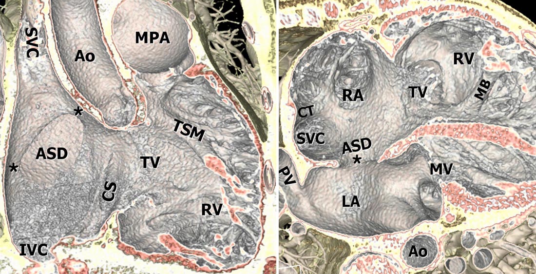
Introduction: Although volume rendering 3-dimensional image using an endo-luminal casting model is a robust method for the evaluation of great vessels, it was rarely used to assess the intracardiac anomaly. We have applied endocardiovascular volume rendering for the intracardiac anomaly. The aim of this study was to introduce the key steps of the technique and to access its feasibility and usefulness.
Methods: Using the 64-slice MD CT data, we underwent endocardiovascular volume rendering in 42 adult patients with congenital heart disease. Unlike traditional endo-cast rendering of enhanced lumen, the endocardiovascular rendering was performed by applying trapezoids of transfer function to depict the cardiac wall and the surrounding soft tissue. We encoded different colors for fat and cardiac muscle and dissected out the obscuring near wall to disclose the internal anatomy of interest. One radiologist and cardiologist assessed the image quality and usefulness of information.
Results: Indications of CT were mostly ASD to evaluate a rim deficiency before device closure in 31 patients, and others were VSD to evaluate complications during the natural course in 3, unroofed coronary sinus in 2, sinus venous ASD in 1, and PDA in 4. The overall image quality was either good or excellent in 76% of cases. Eight patients showed poor image quality due to speckling artifacts by unopacified blood from the caval veins. It provided accurate sizing of ASD and straightforward information about the relationships of the defect and adjacent structures in 24 cases (57%). Rim deficiency was noted mostly at antero-superior rim in 74%.
Conclusion: Virtual dissection CT angiography with endocardiovascular rendering offered comprehensive information about intracardaic anomaly and diagnostic image quality in adult patients with congenital heart disease, especially in the ASD patients. It provided straightforward and easily understandable intracardiac anatomy free from obscuring structures for the interventionist and the cardiac surgeon.
|
|
|
Warning: getimagesize(/home/virtual/circulationadmin/renewal/econgress/conference/abstract/img_files/Figureall2.jpg) [function.getimagesize]: failed to open stream: No such file or directory in /home/virtual/circulationadmin/new/econgress/conference/manage/schedule/view_abstract.php on line 164
|
|


