| єя«•«ьљƒ : ∆чљЇ≈Ќ
|
ЅҐЉцєш»£ - 540409 53 |
| Relationship between fractional flow reserve and coronary angiographic and intravascular ultrasound parameters in ostial lesions: Major coronary ostial lesions vs. Side branch ostial lesions
|
| мДЬмЪЄлМАнХЩкµРл≥СмЫР мИЬнЩШкЄ∞лВік≥Љ¬є,мХДм£ЉлМАнХЩкµР л≥СмЫР¬≤,к≥Дл™ЕлМАнХЩкµР лПЩмВ∞л≥СмЫР¬≥, мЭЄм†ЬлМАнХЩкµРл∞±л≥СмЫРвБі |
| к≥†мІДмЛ†¬є, кµђл≥ЄкґМ¬є,мЦСнШХл™®¬≤,лПДм§АнШХвБі,л∞Хк≤љмЪ∞¬є,к∞ХнШДмЮђ¬є,лВ®м∞љмЪ±¬≥,нЧИмКєнШЄ¬≥,кєАнЪ®мИШ¬є,нГБмКєм†Ь¬≤,мШ§л≥СнЭђ¬є,л∞ХмШБл∞∞¬є |
Background: Angiographic evaluation for ostial lesions is reported to be inaccurate in the assessment of the functional and clinical significance of a lesion.
Objectives: We performed this study to determine the relations between coronary angiography(CAG), intravascular ultrasound (IVUS) and fractional flow reserve (FFR) in coronary ostial lesions.
Methods: CAG, IVUS and FFR measurement were performed in 52 ostial lesions (21 major coronary artery lesions, 31 side branch ostial lesions). Patients with significant proximal or distal lesions other than ostial lesions were excluded. Functionally significant stenosis was defines as FFR <0.8. Hyperemia was induced by intracoronary bolus administration or intravenous infusion of adenosine.
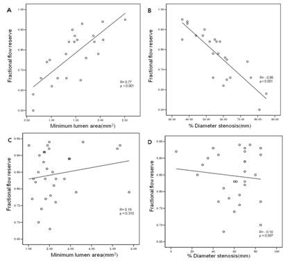
Results: In major coronary artery ostial lesions, there was a positive correlation between FFR and minimum lumen area (MLA) by IVUS (r=0.77, p<0.0001.Fig.A) and a negative correlation between FFR and angiographic % diameter stenosis (r=-0.85, p<0.0001 Fig.B). However, in side branch ostial lesions, there was no correlation between FFR vs. MLA by IVUS and FFR vs. angiographic % diameter stenosis(Fig.C,D). Percent area stenosis also does not have correlation with FFR. Among lesions with MLA <2.0mm2 (n=16), only 25% of the lesions were functionally significant.
Conclusions: In ostial lesions, the relationship between angiographic/IVUS parameters and FFR were different between major coronary artery lesions and side branch lesions.
Figure. Comparison of minimum lumen diameter/area and fractional flow reserve in ostial lesions of major coronary arteries (A,B) and side branches (C,D).
|
|
|
Warning: getimagesize(/home/virtual/circulationadmin/renewal/econgress/conference/abstract/img_files/FFR-7.jpg) [function.getimagesize]: failed to open stream: No such file or directory in /home/virtual/circulationadmin/new/econgress/conference/manage/schedule/view_abstract.php on line 164
|
|


