Background: Pulse wave velocity (PWV) has been known to be useful for the evaluation of arterial stiffness. However, it may represent only global characteristics rather than local vascular alterations. We hypothesized that local vascular properties of carotid artery assessed with velocity vector imaging (VVI) would be more sensitive in the detection of early vascular aging compared with PWV.
Methods: We evaluated 81 healthy, normotensive volunteers (36 male) aged from 20 to 68 years. Transverse images of left common carotid arteries were obtained and divided into six segments. The peak circumferential strain and strain rate of the six segments were analyzed using VVI and standard deviation (SD) of the time to peak circumferential strain and strain rate of the six segments, which represent synchronicity of arterial expansion during systole, were calculated.
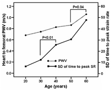
Results: PWV and VVI parameters show significant changes with age. However, the age-related changes in PWV and VVI parameters were different. PWV was increasing with age (r=0.62, p<0.01), but significant increase was observed at the age of 60 (age<60: 767¬±127 cm/s vs ageвЙ•60: 1058¬±287 cm/s, p<0.01). Strain and strain rate of carotid artery decreased significantly with age (r=-0.61, p<0.01; r=-0.68, p<0.01), especially after the age of 40. SD of time to peak strain and strain rate were increasing with age (r=0.58, p<0.01; r=0.70, p<0.01), suggesting dyssynchronous expansion of carotid artery during systole. Importantly, the changes in SD of time to peak strain and strain rate started even in younger age and increased gradually with age.
Conclusion: Arterial assessment using VVI may represent a new method for vascular assessment noninvasively. Dyssynchronous arterial expansion is a sensitive marker of early arterial aging while PWV would be useful at a ripe old age.
Figure. Pulse wave velocity and SD of time to peak circumferential strain rate according to age.
|