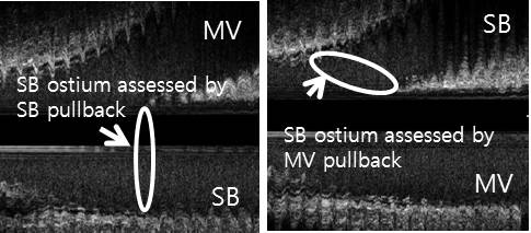
Background Intravascular ultrasound (IVUS) evaluation is helpful in treating bifurcation lesions. However, the axis of IVUS catheter is not always aligned with the axis of artery and simple evaluation of two-dimensional cross section image of side branch ostium may be misleading. Methods From our IVUS database, patients were included if both main vessel (MV) and side branch (SB) pullback in the same procedure were done before procedure. We obtained images of side branch ostium in two different pull-back directions. SB ostium assessed with SB pullback was obtained using cross section image at the site of carina tip. SB ostium assessed with MV pullback was reconstructed using 10 cross sections containing SB ostium (figure). Results 24 lesions were analyzed in this study. 18 lesions were located at distal left main artery and the others were located at LAD (n=4) and LCx (n=2) arteries. SB ostium assessed with SB pullback showed significantly smaller EEM area (13.6±4.7mm2 vs. 16.2±4.0mm2, p<0.001) and plaque & media area (7.2±2.0mm2 vs. 11.7±3.1mm2, p<0.001) compared with MV pullback. On the contrary, lumen area of SB ostium assessed with SB pullback was significantly larger (6.4±3.1mm2 vs. 4.5mm2, p<0.05) and plaque burden (52.9±12.1% vs. 72.2±16.5%, p<0.005). Lumen was more oval and plaque was more eccentric with SB pullback. Conclusions IVUS evaluation of SB ostium by SB pullback underestimates the degree of stenosis.
|