| ЙпЧЅЧќНФ : ЦїНКХЭБИПЌ
|
СЂМіЙјШЃ - 550056 16 |
| Strain Imaging Diastolic Index is a Useful Marker of Diastolic Dysfunction |
| ь ыЈыэъЕыГь ьЌьЅьМэАТЙ , ыЏИъЕ ыЉьДьЄэДыІЌы ьЌьЅыДъГМТВ |
| ъЙъГэТЙ , ыАьЂ
ьЖТЙ, ьЄэьЃМТЙ, ьЄыЈьТЙ, эььЄТЙ, ыАэьБТЙ, ъЙьЃМэТЙ, ььъЗМТЙ, ь ыЊ
эИТЙ, ьЁАь ъДТЙ, ъАь ьБТЙ, ьЄьЌъБДТВ |
Background: Exercise induced delayed diastolic relaxation of myocardial strain, strain imaging diastolic index (SI-DI) after exercise, is known to be a sensitive and reliable marker for myocardial ischemia. We hypothesized that the diastolic relaxation of myocardial strain might be delayed in patients with diastolic heart failure than in normal controls, even in the resting state. Therefore, the aim of the present study was to investigate the usefulness of SI-DI measurements in the evaluation of diastolic dysfunction.
Methods: SI-DI was measured in 20 controls and 20 cardiac amyloidosis (CA) patients with DHF and normal systolic function. SI-DI was determined as (end-systolic strain value - strain value at the one-third of diastolic duration)/(end-systolic strain value) x 100% and compared.
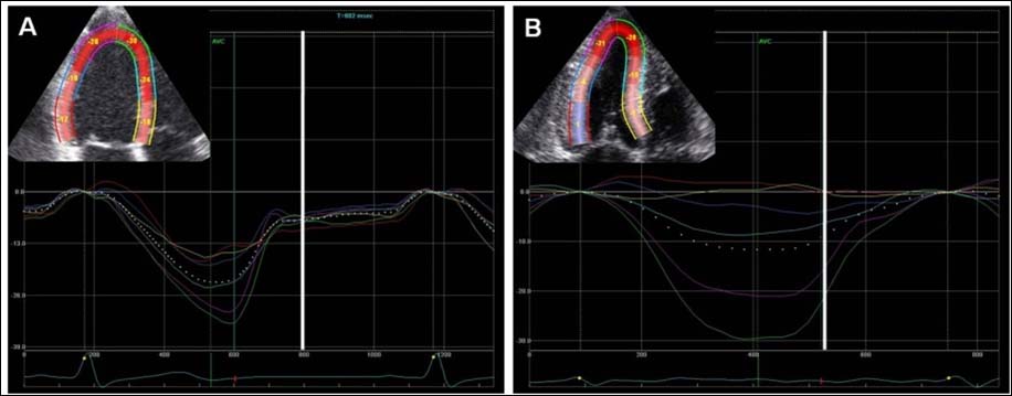
Results: The strain value and SI-DI of each segment was significantly decreased in patients with CA than in controls. The end-systolic strain value of all segments was significantly decreased in patients with CA than that of controls (-20.7ТБ2.7 vs -12.3ТБ5.4%, p<0.001). SI-DI of all segments was significantly decreased in patients with CA than in controls (29.4ТБ18.6 vs 68.2ТБ6.6%, p<0.001). SI-DI of the segments with normal systolic contraction (strain value < -17%) was also significantly decreased in patients with CA than in controls (42.7ТБ8.6 vs 75.5ТБ7.4%, p<0.001) (Figure 1).
Conclusion: Despite of normal systolic function and contraction, diastolic relaxation measured by SI-DI was significantly delayed in patients with DHF by CA than in controls. SI-DI can be a useful new echocardiographic parameter in the differential diagnosis of diastolic function, especially normal diastolic function from pesudonormalization.
|
|
|
Warning: getimagesize(/home/virtual/circulationadmin/renewal/econgress/conference/abstract/img_files/Figure1-SIDI.jpg) [function.getimagesize]: failed to open stream: No such file or directory in /home/virtual/circulationadmin/new/econgress/conference/manage/schedule/view_abstract.php on line 164
|
|


