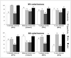
Evaluation of ostial lesion is clinically important as main vessel ostial lesion can cause ischemia in large myocardial territory and ostial lesions usually require complex interventions. However, previous study showed that the angiographic evaluation is not accurate in the prediction of functionally significance in ostial lesions. Moreover, recent IVUS studies suggested that IVUS parameters have limitations in predicting the functional significance of a stenosis. Therefore, we performed this study to assess the relationship of coronary angiography, IVUS and fractional flow reserve (FFR) between major epicardial vessel (MV) and side branch (SB) ostial lesions.107 ostial stenosis of intermediate degree on coronary angiography and underwent both IVUS and FFR were prospectively enrolled in this study. Optimal angiographic and IVUS criteria and their diagnostic accuracy for functionally significant stenoses (FFR вЙ§0.8) were assessed.In MV ostial lesions, FFR had negative correlation with angiographic % diameter stenosis (r=-0.68,p<.001), minimum lumen area (MLA) by IVUS (r=0.55, p<0.001), % plaque burden (r=-0.42,p=0.011) and % area stenosis (r=-0.49,p= 0.003). Meanwhile, FFR had no correlation with angiographic % diameter stenosis (r=-0.067,p=0.635) and weak correlation with MLA (r=0.30,p=0.026) in SB ostial lesions. In MV ostial lesions, best cutoff value of angiographic % diameter stenosis, MLA, % plaque burden and % area stenosis to determine the functional significance was 53%, 3.5mm2, 70% and 50%. However, statistically significant cutoff value of % diameter stenosis and MLA could not be found in SB ostial lesions. Diagnostic performance of IVUS and angiographic parameters was better in MV lesions than in SB ostial lesions (figure).The relations between angiographic/IVUS parameters and FFR were different between MV and SB ostial lesions. Angiographic and IVUS parameters had good correlation with FFR in MV ostial lesions. However, these parameters had poor diagnostic accuracy in predicting the functional significance of SB ostial lesions.
|