| єя«•«ьљƒ : ±Єњђ
|
ЅҐЉцєш»£ - 550432 122 |
| Comparison between fractional flow reserve versus visual assessment and quantitative angiography for coronary artery stenosis in unselected patient cohort. |
| ¬є мДЬмЪЄлМАнХЩкµРл≥СмЫР мЛђнШИкіАмДЉнД∞, ¬≤ лґДлЛємДЬмЪЄлМАнХЩкµРл≥СмЫР, ¬≥ мДЬмЪЄлМАнХЩкµРл≥ілЭЉлІ§л≥СмЫР |
| мЭімГБмЦЄ¬є , кµђл≥ЄкґМ¬є,Todung Silalahi¬є , Hardjo Prawira¬є , кєАмІАнШД¬є , к≥†мІДмЛ†¬є ,мЦСнХЬл™® ¬є ,л∞Хк≤љмЪ∞ ¬є , к∞ХнШДмЮђ¬є ,м°∞мШБмДЭ ¬≤, мЧ∞нГЬмІД ¬≤ , м†ХмЪ∞мШБ¬≥ , кєАмГБнШД¬≥ , м±ДмЭЄнШЄ¬≤ , мµЬлПЩм£Љ¬≤ , кєАнЪ®мИШ¬є , мШ§л≥СнЭђ¬є , л∞ХмШБл∞∞¬є |
Background: Though fractional flow reserve (FFR) can determine the physiologic significance of a stenosis and help ischemia-directed revascularization, anatomic assessment of lesion severity has been the standard for determination of clinically indicated revascularization in daily practice and clinical trials.
Objectives: We investigated the value of the anatomic lesion severity measured by visual assessment or quantitative coronary angiographic (QCA) for predicting the functional severity assessed by FFR in unselected consecutive patient cohort.
Methods: FFR was measured in 872 de novo lesions of 584 patients before revascularization. FFR measured at in-stent restenosis, side branches and graft vessels were excluded. FFR вЙ§0.8 was considered significant. Visual assessment and QCA was performed in an independent core laboratory blinded to the results of FFR.
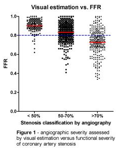
Results: Among the 872 lesions, 49 (5.6%) lesions were at left main (LM), 447 (51.3%) at left anterior descending (LAD), 209 (24%) at left circumflex artery (LCx), 167 (19.2%) at right coronary artery (RCA). 238 (27.3%) lesions were at the vessels sized вЙ•3mm. FFR averaged 0.81¬±0.12 and was вЙ§0.80 in 346 (39.7%) lesions. By QCA, there was weak inverse linear correlation between diameter stenosis and FFR (r=-0.370, p<0.001). Receiver operating curves of QCA to predict FFR вЙ§0.80 revealed C-statistics of diameter stenosis 0.687 and cut-off value 55.5% with sensitivity 0.67 and specificity 0.64. C-statistics of diameter stenosis in the lesions with vessel diameter вЙ•3mm were 0.717. C-statistics was especially low in LM and LCx lesions, 0.580 and 0.659, respectively. By visual assessment, 130/139 (93.5%) lesions were functionally insignificant with stenosis less than 50%, 318/469 (67.8%) with 50% to 70% stenosis, and 75/255 (29.4%) with 71% to 99% stenosis (Figure).
Conclusion: Anatomic lesion severities evaluated by visual assessment and QCA can not accurately predict the functional severity assessed by FFR in unselected large patient cohort.
|
|
|
Warning: getimagesize(/home/virtual/circulationadmin/renewal/econgress/conference/abstract/img_files/fig_550432.jpg) [function.getimagesize]: failed to open stream: No such file or directory in /home/virtual/circulationadmin/new/econgress/conference/manage/schedule/view_abstract.php on line 164
|
|


