| мЖ°мҐЕлѓЉ¬є , мЛђнГЬмД†¬≤ , мµЬмДЭмЫР¬є , мµЬнШХмШ§¬є , кєАмЪ©кЈ†¬є , м†ХмД±нШЄ¬≥ ,кєАмД±нХЬвБі, кєАлМАнЭђ ¬є , к∞ХлНХнШД¬є , мЖ°мЮђкіА¬є |
Background: The role of the Myocbacterium tuberculosis–specific enzyme-linked immunosorbent spot (ELISPOT) assay for diagnosis of tuberculous pericardial effusion (TPE) has not been evaluated.
Methods: We prospectively enrolled consecutive 38 patients with significant pericardial effusion (PE) of unknown etiology. ELISPOT were performed on blood and PE fluid using Mycobacterium tuberculosis–specific antigens (ESAT-6 and CFP-10), and adenosine deaminase (ADA) and interferon gamma (INT-ќ≥) levels in PE were measured. Two definite and 9 probable TPE cases were confirmed by previously published criteria.
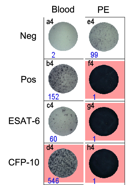
Results: ROC curve analyses showed that areas under curves for diagnosing TPE were 0.748 and 0.691 for ќФ(ESAT-6 - negative control (NC)) and ќФ(CFP-10 - NC) values obtained from blood, while they were 0.946, 0.744, 0.854 and 0.838 for ADA, INT-ќ≥, ќФ(ESAT-6 - NC) and ќФ(CFP-10 - NC) levels derived from the PE fluid. Sensitivities and specificities for diagnosing TPE were 91% and 82% by ADA level (вЙ•40 U/L), 62% and 100% by INT-ќ≥ (вЙ•200 pg/L), 90% and 85% by ќФ(ESAT-6 - NC) (вЙ•49), and 90% and 73% by ќФ(CFP-10 - NC) (вЙ•4) from PE fluid, respectively. For diagnosing definite TPE, ROC curve analyses showed that areas under curves were 0.564, 0.814 for ќФ(ESAT-6 - NC), ќФ(CFP-10 - NC) values obtained from blood, while they were 0.968, 0.946, 0.971, and 0.971 for ADA, INT-ќ≥, ќФ(ESAT-6 - NC) and ќФ(CFP-10 - NC) levels derived from the PE fluid. Sensitivities and specificities for diagnosing definite TPE were 100% and 93% by ADA level (вЙ•95 U/L), 100% and 93% by INT-ќ≥ (вЙ•400 pg/L), 100% and 97% by ќФ(ESAT-6 - NC) (вЙ•1200), and 100% and 97% by ќФ(CFP-10 - NC) (вЙ•1250) from PE fluid, respectively. In all patients with definite TPE, markedly increased number of sensitized T cells to both ESAT-6 and CFP-10 were found in PE (Figure).
Conclusions: ELISPOT assay using PE fluid has a comparable diagnostic power with ADA or INT-ќ≥ for identifying TPE, especially for definite TPE which shows a unique ELISPOT finding.
|