Aim To evaluate angiographic patterns of restenosis following implantation of 2nd generation DES in comparable unselected lesions.
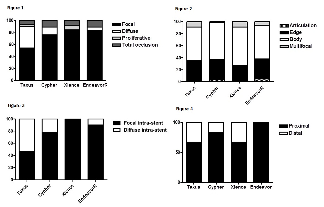
Methods We have identified 242 episodes of restenosis after DES implantation in our institutions between May 2003 and December 2010. Restenosis pattern was classified as focal, diffuse, proliferative, or occlusive. The position of focal restenosis was also categorized as articulation, edge, body, or multi-focal. We have characterized 117, 92, 13 and 20 restenotic lesions in Taxus, Cypher, Xience and EndeavorR stent groups, respectively.
Results For 2nd generation DES, focal restenosis was much higher than 1st generation DES (85% vs 65%, P<0.001). In contrast, just under half of restenosis in Taxus stent was the more severe non-focal pattern (Figure 1). There was no difference in the patterns of focal restenosis between 1st and 2nd generation DES ISR. Edge restenosis was not increased in 2nd generation DES (Figure 2). For intra-stent restenosis, however, diffuse restenosis was significantly decreased and focal restenosis was increased in 2nd generation DES (Figure 3). Multivariable analysis confirmed the positive association of Taxus stent, hypertension, lesion length and stent diameter with non-focal restenosis (Table). The majority of focal restenosis occurs at the proximal stent margin (Figure 4).
Conclusions Focal restenosis was the most common pattern in 2nd generation DES ISR. Diffuse intra-stent restenosis was decreased, but there was no change in the incidence of edge restenosis.
|