| єя«•«ьљƒ : њђ±ЄЇсєя«•
|
ЅҐЉцєш»£ - 550798 249 |
| FDG carotid PET/CT can depict the inflammation on echolucent carotid plaque |
| к∞АнЖ®л¶≠ мЭШк≥ЉлМАнХЩ мИЬнЩШкЄ∞ лВік≥Љ¬є,нХµмЭШнХЩк≥Љ ¬≤ |
| мµЬмЬ§мДЭ ¬є , мЬ§нШЄм§С ¬є,мЬ§м†ХмИЩ ¬є, м†ХмЪ∞л∞± ¬є,л∞Хм≤†мИШ ¬є,мЭілІМмШБ ¬є,м†ХмЪ±мД± ¬є,мКєкЄ∞л∞∞ ¬є,м†ХмЪ©мХИ ¬≤ |
Objectives: To elucidate the relation between the echolucent plaque on carotid ultrasound and acute inflammation on F-18 FDG carotid PET/CT
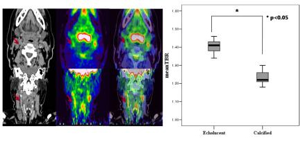
Methods: Thirty two patients (M:F ratio =19:13, mean age=62 ¬± 13 years) who underwent carotid ultrasound were divided into two groups—echolucent plaque (n=22), calcified (n=10). All of the patients underwent F-18 FDG carotid PET/CT. The mean standardized uptake values (SUV), namely target to background ratio (TBR) on 180 min delayed F-18 FDG carotid PET/CT images were compared with serum level of secretory phospholipase A2(sPLA2, Cayman Chemical co., Ann Arbor, MI, USA) in terms of the presence of carotid plaque on carotid US.
Results: 180 min TBR of carotid arterial wall at echolucent plaque, calcified plaque were 1.40±0.05, 1.23±0.03 in both carotid artery. TBR of carotid arterial walls for echolucent plaque were significantly larger than TBR for calcified plaque at the both side of carotid artery (p<0.05)(Fig). sPLA2 levels in echolucent was larger than in calcified plaque but did not reach the statistical significance.(1.12±1.29 vs 1.04±1.32 ng/ml, p=0.07)
Conclusions: Our results show that F-18 FDG carotid PET/CT can depict metabolically active echolucent carotid plaques. Furthermore secretory phospholipase A2 need to be inverstigated for surrogates of atherosclerotic plaque
|
|
|
Warning: getimagesize(/home/virtual/circulationadmin/renewal/econgress/conference/abstract/img_files/fig_550798.jpg) [function.getimagesize]: failed to open stream: No such file or directory in /home/virtual/circulationadmin/new/econgress/conference/manage/schedule/view_abstract.php on line 164
|
|


