| єя«•«ьљƒ : ∆чљЇ≈Ќ
|
ЅҐЉцєш»£ - 550918 88 |
| Semi-quantitative Visual Analysis of Myocardial Perfusion Scitigraphy Is Helpful for Differential Diagnosis of Bundle Branch Block and Myocardial Infarction |
| к∞АнЖ®л¶≠лМАнХЩкµР мИЬнЩШкЄ∞лВік≥Љ¬є, мШБмГБмЭШнХЩк≥Љ¬≤, нХµмЭШнХЩк≥Љ¬≥ |
| мµЬкЈЬмШБ¬є , м°∞мЭАм£Љ¬є, кґМл≤Фм§А¬є, кєАлПЩлєИ¬є, мЮ•мД±мЫР¬є, л∞Хм∞ђмДЭ¬є, м†ХнХімЦµ¬є, м†ДнЭђк≤љ¬є, мЬ§нШЄм§С¬є, кєАмЮђнШХ¬є, мЭіл∞∞мШБ¬≤ , мШ§м£ЉнШД¬≥ |
Background: It has been suggested that even without ischemia or infarction, left bundle branch block (LBBB) itself may reduce myocardial perfusion. In this study, we aimed to evaluate whether semi-quantitative visual analysis of perfusion defect could discriminate myocardial infarction (MI) from BBB without significant coronary artery disease.
Method: The 79 patients with BBB (BBB group, mean age=68±9 years, 45 males, RBBB (n) = 39 and LBBB (n) = 40) who showed fixed perfusion defect without significant coronary artery stenosis or 59 patients with history of MI (post MI group, mean age=65±9 years, 42 males) were evaluated. All subjects had taken ECG gated stress-rest SPECT using 99mTc compounds and coronary angiography. A segmentation model has been standardized for semi-quantitative visual analysis by dividing the myocardium into 20 segments in perfusion scintigraphy. The sum of the segmental scores from the stress images (stress score) and the rest images (rest score) were calculated and compared between both groups.
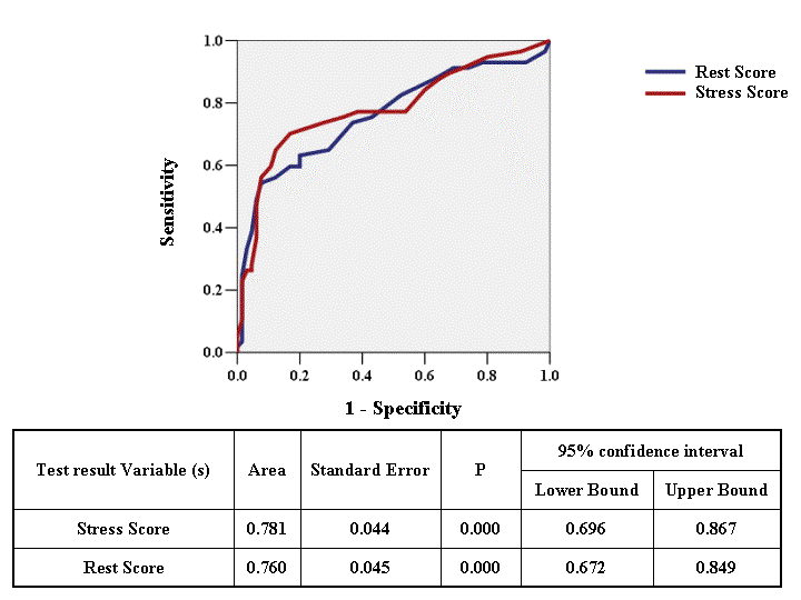
Results: There were no significant differences in systolic function (ejection fraction) between both groups (52±11% for BBB vs. 47±13% for post MI, p=0.54). However, the % thickening (relative change in thickening of myocardium during systole) analyzed in myocardial scintigraphy was significantly low in post MI patients compared with BBB patients. Also, the post MI patients showed significantly high rest score (8.3±5.4 vs. 15.0±8.1, p<0.001) and stress score (10.7±6.0 vs. 18.4±8.4,p<0.001) compared with BBB patients. With a cut off value of stress score 14.5, post MI perfusion defect could be predicted (74% of sensitivity and 73% of specificity, Figure).
Conclusion: Segmental perfusion defect score by visual analysis of myocardial scintigraphy is significantly high in MI group compared with BBB group. Semi-quatitative measurement of stress score in myocardial perfusion scintigraphy is feasible to differentiate between MI and BBB in patient with positive perfusion defect.
|
|
|
Warning: getimagesize(/home/virtual/circulationadmin/renewal/econgress/conference/abstract/img_files/ROCcurveforMIandBBB.gif) [function.getimagesize]: failed to open stream: No such file or directory in /home/virtual/circulationadmin/new/econgress/conference/manage/schedule/view_abstract.php on line 164
|
|


