Intervention of the Month

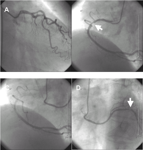
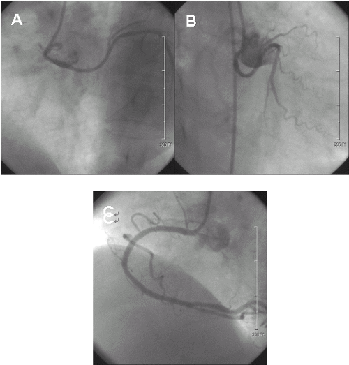
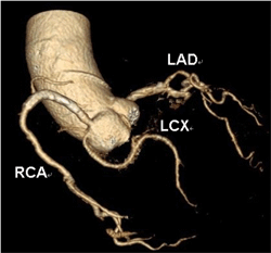
Successful Coronary Intervention in a Patient with Separate Origin of Three Coronary Arteries
JYoungkeun Ahn, MD, PhD, FACC, FSCAI, Young Joon Hong, MD, PhD, Yun Hyeon Kim, MD, PhD, and Myung Ho Jeong, MD, PhD, FACC, FAHA, FESC, FSCAI
Departments of Cardiovascular Medicine and Radiology, Chonnam National University Hospital, Gwangju, Korea

A 69-year-old woman with a 7-year history of treated essential hypertension presented with a one-year history of effort angina and dyspnea. Her resting electrocardiogram showed left ventricular hypertrophy and ejection fraction by echocardiogram was 71%. Her coronary angiogram revealed complete separation of three coronary ostia and critical stenosis in proximal right coronary artery and proximal left circumflex artery (Figure 1). To the best of our knowledge, this particular combination of anomalies has not been reported previously. Taxus stent (3.0*32 mm) was deployed for proximal right coronary artery and balloon angioplasty using 2.5mm balloon was performed for proximal left circumflex artery. Final coronary angiogram showed no residual stenosis with good distal flow over right coronary artery and left circumflex artery (Figure 2). Computed tomographic coronary angiography with retrospective electrocardiogram gated 64-slice multi-detector demonstrated the separate origin of three coronary arteries (Figure 3). Six months follow-up coronary angiogram demonstrated no restenosis in right coronary artery and left circumflex artery.

¡ã Figure 1.Coronary angiogram showing anomalous origin of coronary arteries. A: Left coronary angiogram (right anterior oblique caudal view) showed left anterior descending artery (LAD) but no visualization of left circumflex artery (LCX). B: Right coronary angiogram (left anterior oblique view) showed right coronary artery (RCA) with critical stenosis in proximal portion. C: The LCX is originated from a separate ostium near the ostium of RCA. This figure was obtained shortly after dye filling of RCA. D: The LCX was visualized from a separate ostium. White arrows indicate stenosis.
 

¡ã Figure 2.Coronary angiogram after balloon angioplasty for proximal left circumflex artery (A; left anterior oblique view, B; right anterior oblique caudal view) and stenting for proximal right coronary artery (C).
 

¡ã Figure 3.Computed tomographic coronary angiography demonstrated the separate origin of three coronary arteries (LAD; left anterior descending artery, LCX; left circumflex artery, RCA: right coronary artery).


À§ÀÇ ÀÌ´ÞÀÇ ÁßÀç¼úÀ» º¸½Å ¼±»ý´ÔµéÀÇ ÀǰßÀ» ¾Æ·¡ÀÇ ÀÇ°ß ¾²±â¿¡ ÀǰßÀ» ³²°Ü ÁֽʽÿÀ.
¼±»ý´ÔµéÀÇ ´Ù¾çÇÑ ÀǰßÀ» ¹Þ½À´Ï´Ù.

[Àǰ߾²±â]