 Intervention of the Month Intervention of the Month |
|
| Recurrent Stent Thrombosis and Pulmonary Thromboembolism Associated With Hyperhomocysteinemia |
Min Chul Kim, MD, Kyung Hoon Cho, MD, Daisuke Hachinohe, MD, Khurshid Ahmed, MD, Keun Ho Park, MD, Doo Sun Sim, MD, Young Joon Hong, MD, PhD, Ju Han Kim, MD, PhD, Youngkeun Ahn, MD, PhD, FACC, FSCAI and Myung Ho Jeong, MD, PhD, FACC, FAHA, FESC, FSACI
The Heart Center of Chonnam National University Hospital, Gwangju, Korea
|
|
A 75-year-old male developed sudden chest pain 3 hours ago. Pain lasted during 1 hour and he suddenly collapsed. He was rushed unconscious to a neighborhood hospital by emergency medical services. And then he transferred to our hospital for further evaluation. He was a current smoker at 70 pack-years and no other atherosclerotic risk factors were presented. His initial rhythm was junctional bradycardia with 45 beats per minutes of heart rate and blood pressure was 70/40 mmHg. Also, 12-lead electrocardiogram showed ST-segment elevation in II, III, and aVF and V4R, V5R, V6R, V7, V8, and V9 leads on right posterior electrocardiogram (Fig. 1). Primary percutaneous coronary intervention (PCI) was performed and diagnostic coronary angiography showed thrombotic total occlusion in proximal right coronary artery (RCA) and lesion was successfully revascularized with stenting (Coroflex-blue 4.0x19 mm, B. Braun Systems) (Fig. 2). 2-D echocardiography showed mild LV systolic dysfunction and hypokinesia in RCA territory. During admission, he complained chest discomfort and chest X-ray showed haziness in right lung field. Chest computed tomography (CT) scan revealed filling defects in segmental pulmonary arteries in left lower lobe (Fig. 3). Although RV dysfunction was presented, his vital sign and general condition were stable. We performed only anticoagulation without thrombolysis. He was discharged with triple ant-platelet therapy on event-free state 7 days later.
After 1 month, he revisited our emergency department with typical angina. Twelve-lead electrocardiogram showed T-inversion in II, III, and aVF. Urgent coronary angiography (CAG) showed thrombosis and de novo lesion in distal part of stent. We carried out direct stenting using bare-metal stent (Coroflex-blue 3.5x19 mm, B. Braun Systems) (Fig. 4). Despite of successful PCI, patient complained chest pain immediately after PCI. 12-lead ECG during development of pain showed STE in II, III, and aVF with complete atrioventricular block. Emergent CAG showed critical spasm over RCA and stent thrombosis. After nitrate and abciximab infusion, distal flow was improved (Fig. 5). He did not have any trouble with medication and no chest pain was developed in other day except for the second admission day.
After discharge with other event, he revisited our hospital 5 months later. However, CAG at that time showed no in-stent restenosis, thrombosis and de novo lesions (Fig. 6). He was discharge and followed up at outpatient department without angina. At 9 months after last admission, he revisited our emergency department with comatose mental status and hypotension. 12-lead ECG showed ST-segment elevation in II, III, and aVF with complete atrioventricular block. Emergent CAG revealed total occlusion of proximal RCA due to recurrent stent thrombosis in proximal RCA and we performed successful PCI with ballooning and final CAG showed TIMI (Thrombolysis In Myocardial Infarction) III antegrade flow with small amount of remnant thrombosis (Fig. 7). During admission, no other cardiovascular events were developed. Platelet resistance test showed no resistance to anti-platelet (aspirin 449 ARU / P2Y12 90 PRU and 35%). But, hyperhomocysteinemia was detected in other tests for detection of thrombotic tendency (19.43 u mol/L). We prescribed folate in addition to triple antiplatet therapy. He ceased warfarin earlier compared with recommended medication period. However, other medication compliance was good (Fig. 8). Although this patient did not stop smoking, this cannot explain all events of stent thrombosis and pulmonary thromboembolism in this patient. Furthermore, patient experienced all types of stent thrombosis including acute, subacute and very late stent thrombosis despite of bare-metal stent implantation. Therefore, we prescribed folate to prevent vascular thrombotic event in addition to triple antiplatelet therapy. There has been controversy whether folate is helpful for prevention of vascular thrombotic event in patient with hyperhomocysteinemia. However, we think it is valuable in patient without other cardiovascular risk factors. It needs large scale randomized control trial to support this issue.
|
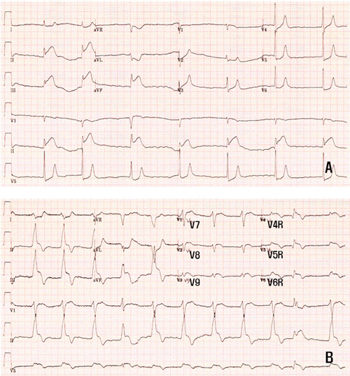 |
▲ Figure 1. ST-segment elevation in II, III, and aVF (Figure 1A) and V4R, V5R, V6R, V7, V8, and V9 leads on right posterior electrocardiogram (Figure 1B)
|
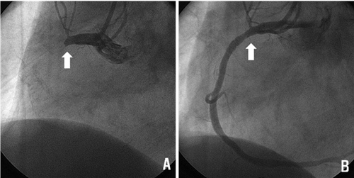 |
▲ Figure 2. Primary PCI; CAG showed TTO in proximal RCA (white arrow) and successful PCI was performed with bare metal stent (Coroflex-blue 4.0x19 mm, B. Braun Systems).
|
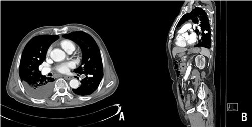 |
▲ Figure 3. Filling defects in segmental pulmonary arteries in left lower lobe (white arrow).
|
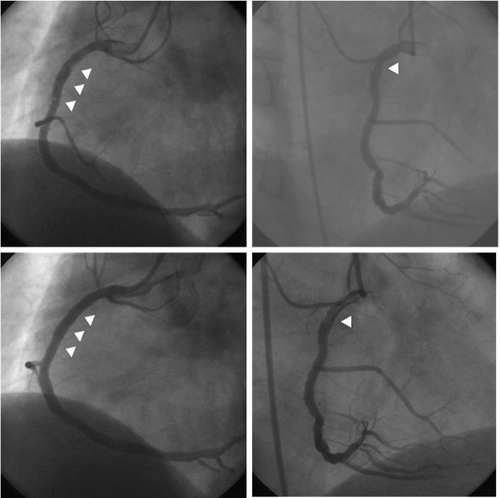 |
▲ Figure 4. Stent thrombosis and de novo lesion in distal to stent (upper two panels, arrow heads). Successful PCI with direct stenting using bare-metal stent (lower two panels, arrow heads, Coroflex-blue 3.5x19 mm, B Braun Systems)
|
 |
▲ Figure 5. Critical spasm over RCA and acute stent thrombosis (Fig. 5A, white arrow). Improved flow coronary flow after nitrate and abciximab infusion (Fig. 5B).
|
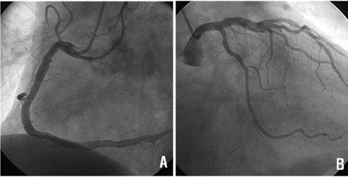 |
▲ Figure 6. Follow-up coronary angiogram after 6 months until first admission day (Figure 6A: Right coronary artery, Figure 6B: Left coronary artery). There were no in-stent restenosis, stent thrombosis, and de novo lesions.
|
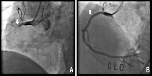 |
▲ Figure 7. Stent thrombosis in proximal RCA stent (Fig. 7A). Successful primary PCI was performed with the aid of intracoronary glycoprotein IIb/IIIa inhibitor and there were small amount of remnant thrombi in culprit lesion (Fig. 7B).
|
 |
▲ Figure 8. Duration of anti-platelet and oral anti-coagulation medication. His stent thrombosis events; acute, subacute, and very late stent thrombosis are presented in non painted box during clinical follow up perio.d
|
|
| |
위의 이달의 중재술을 보신 선생님들의 의견을 아래의 의견 쓰기에 의견을 남겨 주십시오.
선생님들의 다양한 의견을 받습니다.
|
![[의견쓰기]](/image/comment_btn.gif) |
Warning: include(/home/virtual/circulationadmin/new/info/comment_index.php) [function.include]: failed to open stream: No such file or directory in /home/virtual/circulationadmin/new/info/case/201008/case201008.htm on line 186
Warning: include() [function.include]: Failed opening '/home/virtual/circulationadmin/new/info/comment_index.php' for inclusion (include_path='.:/usr/local/php/lib/php') in /home/virtual/circulationadmin/new/info/case/201008/case201008.htm on line 186
|
|
|