|
 Case Case
|
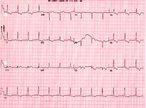
A 35-year-old female was admitted to the hospital because of speech disturbance and right side paresthesia of the body, which gradually worsened. The patient had been well until one month earlier. There was no important finding of family history, physical examination including neurological tests, laboratory data and chest X-ray except the electrocardiographic findings which showed ST segment depression and T wave inversion in lead , aVL, V3-6 and Q wave in V1-2 (Figure 1).
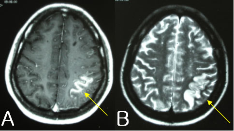
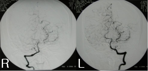
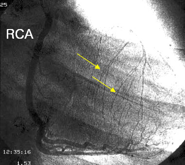
Magnetic resonance imaging of the brain showed infracted area of low signal intensity on the T1-weighted image and high signal intensity on the T2-weighted image in the left superior parietal lobe (Figure 2). A angiography of the both cerebral arteries showed obliterated anterior cerebral arteries and middle cerebral arteries with characteristics "puff of smoke", that is the typical finding of moyamoya disease (Figure 3). Though the patient had no angina history before, a coronary angiography which was performed due to abnormal electrocardiographic findings and hypokinetic wall motion of the anterior myocardium on 2 dimensional echocardiography, showed the left coronary artery with ostial total obstruction and collateral blood flow from normal right coronary artery (Figure 4).
The final diagnosis was moyamoya disease with total occlusion of left main coronary artery.
She had coronary artery bypass graft operation for severe coronary artery disease instead of percutaneous coronary intervention (PCI) because of the left main ostial lesion.
|
Legend
| Fig. 1.Electrocardiography shows ST segment depression and T wave inversion in I, aVL, V3-6 and Q wave in V1-2. |
 |
| Fig. 2. Brain MRI of cerebral infarction with T1-weighted image (arrow in A) and T2-weighted image (arrow in B) in left superior parietal lobe. |
 |
| Fig. 3. Both cerebral angiograms of obliterated anterior cerebral arteries and middle cerebral arteries with characteristics "puff of smoke" or moyamoya apperance |
 |
| Fig. 4. The right coronary angiography showing left coronary artery with ostial total obstruction and collateral blood flow (arrows) from normal right coronary artery. |
 |
☞ 질문이나 의견이 있으시면 회원들의 공간
 에 글을 써주시기 바랍니다 에 글을 써주시기 바랍니다
|