Intervention of the Month Intervention of the Month
|
|
|
|
A Case of Stenting in Guiding Catheter-induced Long Spiral Dissection in the Right Coronary Artery
Young Joon Hong, MD, Weon Kim, MD, PhD, Youngkeun Ahn, MD, PhD, FSCAI, and Myung Ho Jeong, MD, PhD, FACC, FESC, FSCAI
The Heart Center of Chonnam National University Hospital, Gwangju, Korea
|
 |
CASE
|
|
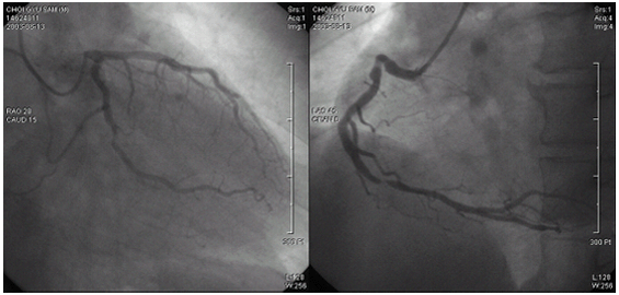
A 57-year-old man visited to Chonnam National University Hospital due to crescendo left anterior chest pain. He was a 30 pack-years current smoker and had a hypertension. His electrocardiogram showed deep T wave inversion more than 3 mm in lead II, III, aVF. The level of cardiac enzyme was normal and the left ventricular ejection fraction on echocardiogram was 73%. A diagnostic coronary angiogram (Figure 1) revealed diffuse insignificant stenosis in middle left anterior descending artery, total occlusion in distal left circumflex artery, and critical eccentric stenosis in proximal and distal right coronary artery (RCA). We decided to perform a percutaneous coronary intervention for stenotic lesions in RCA. After engagement of right guiding catheter, long spiral dissection (type D) was developed over RCA (Figure 2). After guidewire passage over RCA, direct stenting using 3.5*23 mm Bx Sonic stent was performed for proximal RCA. After stenting, final coronary angiogram showed TIMI II flow over RCA. One week after stenting, follow-up right coronary angiogram revealed remained critical stenosis proximal to the previous stented site. Direct stenting using 3.0*16 mm ClearFlex stent was performed for this lesion. After additional high pressure ballooning, final coronary angiogram showed no residual stenosis with good distal flow over RCA.
|
|
|
|
Fig. 1.
Diagnostic coronary angiogram revealed insignificant stenosis in middle left anterior descending artery, total occlusion in distal left circumflex artery, and critical stenosis in proximal and distal right coronary artery.
|
|
|
|
Fig. 2.
After engagement of right guiding catheter, long spiral dissection (type D) was developed over the entire right coronary artery.
|
 |
 |
Fig. 3.
After guidewire passage over right coronary artery, direct stenting using 3.5*23 mm Bx Sonic stent was performed for proximal right coronary artery. After stenting, final coronary angiogram showed TIMI II flow in the distal right coronary artery.
|
|
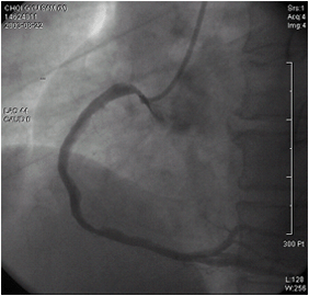 |
Fig. 4.
One week after stenting, follow-up right coronary angiogram revealed remained critical stenosis proximal to the previous stented site.
|
|
|
|
|
Fig. 5.
Direct stenting using 3.0*16 mm ClearFlex stent was performed for proximal right coronary artery proximal to the previous stented site.
|
 |
Fig. 6.
After additional high pressure ballooning, final coronary angiogram showed no residual stenosis with good distal flow over right coronary artery.
|
|
☞ 질문이나 의견이 있으시면 회원들의 공간
 에 글을 써주시기 바랍니다. 에 글을 써주시기 바랍니다. ▲Top
|
|