Echocardiography 3
Novel 4D TTE/TEE Technologies in Valvular Heart Disease

Eun Kyoung Kim, MD., PhD.
Sungkyunkwan Univ, KoreaAdvances in echocardiographic imaging have transformed the assessment and management of valvular heart disease, particularly in the current era of transcatheter structural interventions. The emergence of 4D transthoracic (TTE) and transesophageal echocardiography (TEE) technologies represents a major step forward, offering unprecedented detail in both anatomic reconstruction and hemodynamic evaluation.
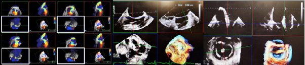
A key innovation is real-time three-dimensional valve reconstruction, now achievable with markedly improved temporal and spatial resolution. High-density volumetric datasets allow clinicians to visualize complex valve morphology from multiple perspectives, providing a "surgeon's view" and "multi-axis view" of the mitral, tricuspid, and aortic valves. These capabilities are invaluable for pre-procedural planning and intra-procedural guidance in transcatheter interventions, where precise delineation of leaflet morphology, commissural orientation, and annular geometry directly impacts device selection, positioning, and procedural success.
Equally important is the application of multiplanar reconstruction (MPR) derived from 4D datasets. By extracting cross-sectional images from 4D datasets, clinicians can interrogate regurgitant orifices and flow convergence zones beyond the constraints of conventional two-dimensional imaging. Through MPR, quantification of regurgitant severity-including direct planimetry of the regurgitant orifice area and refined assessment of vena contracta area-can be performed with improved accuracy and reproducibility. In the context of mitral and tricuspid regurgitation, MPR facilitates integration with volumetric flow methods, thereby advancing quantification from semi-quantitative grading to more robust and physiologically meaningful measurements.
The adoption of these technologies has also enhanced the reproducibility of serial assessments, which is critical for monitoring disease progression, evaluating remodeling, and assessing treatment response. Importantly, 4D echocardiography minimizes reliance on geometric assumptions inherent in 2D methods and artifacts from the implantable devices, providing a more anatomically faithful representation of complex valve pathology, including multi-jet regurgitation and non-circular annular configurations.
As the field advances, 4D TTE/TEE is increasingly moving from research environments into routine clinical practice. The next frontier will involve combining these high-fidelity imaging modalities with computational modeling, artificial intelligence-driven quantification, and multimodality integration, further refining patient selection, procedural guidance, and outcome prediction.