TSOC-KSC Joint Session (Smart Health)
Bridging Cardiology and AI: Innovative Solutions for Coronary Artery Disease Management

Shih-Sheng Chang, MD, PhD
China Medical Univ., TaiwanThe integration of artificial intelligence (AI) into cardiology is transforming how we diagnose, treat, and monitor patients with coronary artery disease (CAD). Over the past decade, advances in deep learning and multimodal data analysis have shifted AI from early research to practical clinical use. These progressions highlight not only technical feasibility but also real-world clinical benefits.
AI-ECG systems represent one of the earliest breakthroughs. By detecting ST-elevation myocardial infarction (STEMI) with area-under-the-curve values above 0.9, AI tools have reduced diagnostic delays and shortened door-to-balloon times. In pre-hospital settings, these systems enable early triage and direct transfer to PCI-capable centers, ultimately improving outcomes in acute coronary syndromes.
In preventive cardiology, deep learning applied to low-dose, non-gated chest CT scans allows opportunistic detection of coronary artery calcium (CAC). Our team developed a two-step model combining nnU-Net and 3D ResNet-18, achieving an overall accuracy of 85.7% and vessel-specific AUROCs above 0.87. This approach transforms routinely acquired lung CTs into powerful predictors of cardiovascular risk, without additional radiation or cost.
Before the cardiac cath procedure, AI-powered clinical decision support tools like ePRISM provide personalized risk assessments for bleeding and contrast-induced nephropathy, helping to lower complications and reduce hospital stays. During coronary interventions, AI enhances both decision-making and safety. AI-derived fractional flow reserve (FFRangio) and AI-based quantitative coronary angiography (AI-QCA) have demonstrated non-inferiority to traditional IVUS or OCT guidance, reducing procedural complexity while maintaining comparable outcomes. Similarly, AI-enhanced intravascular imaging (IVUS and OCT) provides automated lesion assessment with strong correlation to expert analysis, streamlining stent optimization.
Emerging applications go even further. Video-based AI algorithms can now estimate left ventricular ejection fraction directly from routine angiography, providing real-time functional assessment during PCI. This innovation has shown diagnostic accuracy with AUROCs around 0.90, potentially improving safety in urgent or unstable cases.
AI is now a practical partner in every stage of CAD management, from early detection to personalized intervention and long-term care. The next frontier involves integrating hospital systems, obtaining regulatory validation, and building clinician trust with transparent models. These advances help cardiologists progress toward precision medicine, providing CAD patients with timely, tailored, and effective treatments.
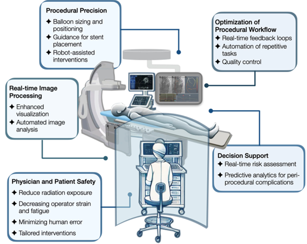
Figure. The "future" cath lab? With AI driving precision, imaging, workflow, and safety, it seems less like tomorrow and more like today. (Adapted from Samant S, et al. J Soc Cardiovasc Angiogr Interv. 2025;4:102519.)