이달의 증례
- 페이지 경로
- HOME > 자료실 > 이달의 증례


|
이달의 중재술 / 2018년 5월 Multivessel disease with recanalized thrombus mimicking coronary dissection: etiologic insights from optical coherence tomography |
||||||
| 저자 | Yongcheol Kim, MD, Myung Ho Jeong, MD, MD, PhD, FACC, FAHA, FESC, FSCAI, Min Chul Kim, MD, PhD, Doo Sun Sim, MD, PhD Young Joon Hong, MD, PhD, Ju Han Kim, MD, PhD and Youngkeun Ahn, MD, FACC, FSCAI | |||||
|---|---|---|---|---|---|---|
| 소속 | The Heart Center of Chonnam National University Hospital, Gwangju, Korea. | |||||
|
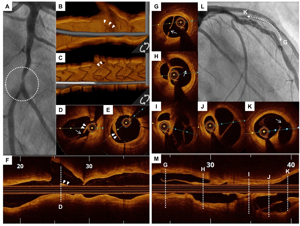
A 65-year-old man with a history of treated diabetes and hypertension presented to our department due to worsening exertional angina. Coronary angiography (CAG) revealed a severe stenosis with haziness in the distal left circumflex artery (LCx) (Panel A, white circle). OCT of the LCx lesion demonstrated diffuse burden with dissection flap (Panels D and F, arrowheads), also confirmed by 3D OCT reconstruction (Panels B, arrowheads), and intimal disruption (Panel D, white arrow), suggestive of plaque rupture. The OCT assessment led to stent implantation with a 2.75 x 18 mm Xience Alpine (Abbot Vascular Santa Clara, CA). Repeated OCT assessment demonstrated excellent stent expansion and good strut apposition with intraluminal tissue prolapse (Panels C and E, arrowheads). Regarding the left anterior descending artery (LAD) lesion, CAG showed multiple linear filling defects with haziness in the mid LAD (Panel L, line with both arrowheads). the OCT with cross-sectional (Panels G to K) and longitudinal view (Panel M) demonstrated a honeycomb-like structure with multiple channels of various sizes, separated by highly backscattering septa, communicating each other (Panels G, H, and K, white arrow.) With this OCT finding of the LAD, we concluded that this characteristic represented recanalized thrombus. The lesion of LAD was treated with 3.0 x 38 mm Xience Alpine and follow-up OCT showed good strut apposition without edge dissection. This case highlights the benefit that the superior resolution (10 µm) of OCT is able to distinguish etiology of intraluminal events, such as recanalized thrombus and plaque rupture, especially in multivessel lesions.
|
||||||
| 첨부파일1 | ||||||

- Tel 02-3275-5258
- Fax 02-3275-5259
- E-mail herz1@circulation.or.kr
- Copyright© The Korean Society of Cardiology


- 심장학 최신지견 따라잡기
- 진료지침
